 English
 Español
NEWS
NEWS
NEWS
NEWS
NEWS
NEWS
NEWS
NEWS
NEWS
NEWS
NEWS
NEWS
NEWS
NEWS
NEWS
NEWS

Peng Shulan — Regional Sales Consultant
Home / Author / Peng Shulan — Regional Sales Consultant / Integrated Cryogenic and Ion-Beam Sample Preparation for High-Fidelity Electron Microscopy
Integrated Cryogenic and Ion-Beam Sample Preparation for High-Fidelity Electron Microscopy
Content
- 1 Why Sample Preparation Determines Electron Microscopy Quality
- 2 Platform Architecture and Product Scope
- 3 LSCT Cryogenic Transfer Systems
- 4 IMP120 Multi-Beam Ion Milling Technology
- 5 HSC Series Etching, Coating, and Milling Systems
- 6 Major Advantages Over Conventional and Fragmented Approaches
- 7 Applications Across Scientific and Industrial Fields
- 8 Integrated Workflow Examples
- 9 Manufacturing and Engineering Strengths
- 10 Operational Benefits for Research and Industry
- 11 Recommended Preparation Considerations
- 12 Competitive Positioning
- 13 Customer Groups and Procurement Value
- 14 Implementation and Training
- 15 Future Development of Electron Microscopy Preparation
- 16 Questions and Answers
- 16.1 What is the main purpose of the Electron Microscopy Sample Preparation Platform?
- 16.2 Which instruments are included in the platform?
- 16.3 Why is cryogenic transfer important?
- 16.4 Can the platform be used for hard materials?
- 16.5 Is the equipment suitable for biological research?
- 16.6 How does ion milling compare with mechanical polishing?
- 16.7 What is the purpose of coating a sample?
- 16.8 Can the systems be customized?
- 16.9 Who are the typical customers?
- 16.10 What should customers prepare before purchasing?
- 17 Conclusion
- 18 References
- 19 Product: Electron Microscopy Sample Preparation Platform
Electron microscopy can reveal structures at the nanometer scale, but the quality of the final image depends heavily on what happens before the sample enters the microscope. A poorly preserved biological specimen may lose its native morphology. A soft polymer may shrink or collapse during preparation. A battery electrode may develop artificial cracks during drying. A semiconductor cross-section may contain redeposited material, curtaining, edge rounding, or thermal damage. In each case, the microscope can only record the condition of the prepared specimen, not the condition of the original sample.
The Electron Microscopy Sample Preparation Platform is designed to address these challenges through an integrated family of cryogenic transfer, ion milling, ion etching, coating, polishing, and sample preparation technologies. The platform includes LSCT series cryogenic transfer systems, the IMP120 multi-beam ion milling system, and HSC series ion beam etching, coating, and milling systems. Working individually or as a coordinated workflow, these instruments help laboratories preserve sample state, improve surface quality, and prepare reliable specimens for scanning electron microscopy, transmission electron microscopy, focused ion beam workflows, and related analytical techniques.
Rather than treating sample preparation as a simple preliminary step, the platform approaches it as a controlled scientific process. Temperature, vacuum, ion energy, beam incidence, surface cleanliness, transfer time, and coating conditions can all influence the validity of microscopy results. By combining controlled cryogenic handling with precision ion-beam processing, the system supports demanding applications in life sciences, materials research, semiconductors, energy storage, pharmaceuticals, geology, and industrial failure analysis.
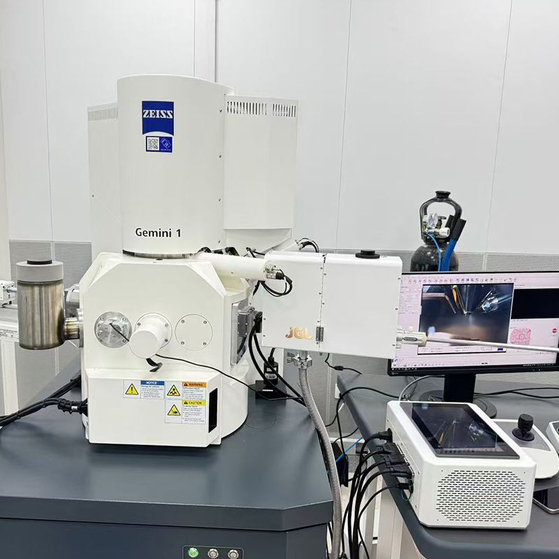
Electron Microscopy Sample Preparation Platform
Why Sample Preparation Determines Electron Microscopy Quality
Electron microscopy requires specimens that are sufficiently thin, stable, clean, and compatible with the electron beam. These requirements vary according to the microscope and the sample, but the underlying principle is consistent: the prepared specimen must represent the structure being studied as accurately as possible.
For biological samples, water is often an essential part of the native structure. Conventional dehydration can cause collapse, extraction of soluble components, membrane distortion, and changes in spatial relationships. Rapid cryogenic fixation and low-temperature transfer help immobilize water and cellular components in a condition closer to the original hydrated state. Maintaining that state during transfer is equally important. If a frozen sample is exposed to moisture or allowed to warm significantly, crystalline ice and contamination may obscure the structure.
Soft materials present a related problem. Polymers, gels, emulsions, oils, and composite materials can deform under vacuum, electron-beam exposure, or mechanical preparation. Cooling the sample reduces mobility and can help stabilize fragile features. It also limits some forms of beam-induced damage and makes it possible to examine interfaces that might otherwise change during preparation.
Hard materials require a different type of control. Metals, ceramics, semiconductors, minerals, battery electrodes, and solid electrolytes may need a flat cross-section, a clean interface, or a precisely thinned region. Mechanical cutting and polishing can introduce scratches, deformation layers, embedded abrasives, burrs, and residual stress. Ion-beam processing provides a non-contact method for removing material and refining the exposed surface, with process parameters selected according to the composition and sensitivity of the specimen.
These examples show why a single preparation technique cannot meet every microscopy requirement. High-quality analysis often requires a sequence of operations: fixation, freezing, transfer, cutting, milling, polishing, coating, observation, and sometimes additional in-situ treatment. An integrated platform reduces the risk of losing sample integrity between these stages.
Platform Architecture and Product Scope
The platform is built around three complementary equipment groups. LSCT series systems provide cryogenic and vacuum transfer. The IMP120 multi-beam ion milling system supports controlled polishing, thinning, and processing across a broad range of sample types. HSC series systems extend the preparation capability through ion beam etching, coating, and milling under ambient or cryogenic conditions.
Equipment group |
Primary function |
Typical contribution to the workflow |
LSCT series |
Cryogenic and vacuum transfer of frozen specimens |
Preserves low-temperature conditions and reduces ice or contamination exposure during transfer |
IMP120 multi-beam ion milling system |
Precision ion milling, polishing, thinning, and cutting |
Creates clean, flat, and controlled surfaces for high-resolution analysis |
HSC series |
Ion beam etching, coating, milling, and related sample treatment |
Supports surface preparation, conductivity enhancement, cross-section work, and cryogenic processing |
The value of the platform lies not only in the capabilities of each instrument, but also in how the instruments support a continuous preparation strategy. A sample can be processed under suitable thermal and vacuum conditions, moved between preparation stages with lower exposure to contamination, and treated according to the requirements of the final microscopy method.
For example, a frozen biological specimen may be cryogenically transferred from a preparation environment to an electron microscope without unnecessary warming. A battery electrode may be stabilized at low temperature, opened or polished with an ion beam, and then transferred for examination of the electrode-electrolyte interface. A semiconductor device may undergo controlled milling to expose a failure site while minimizing mechanical deformation and unwanted material redeposition.
LSCT Cryogenic Transfer Systems
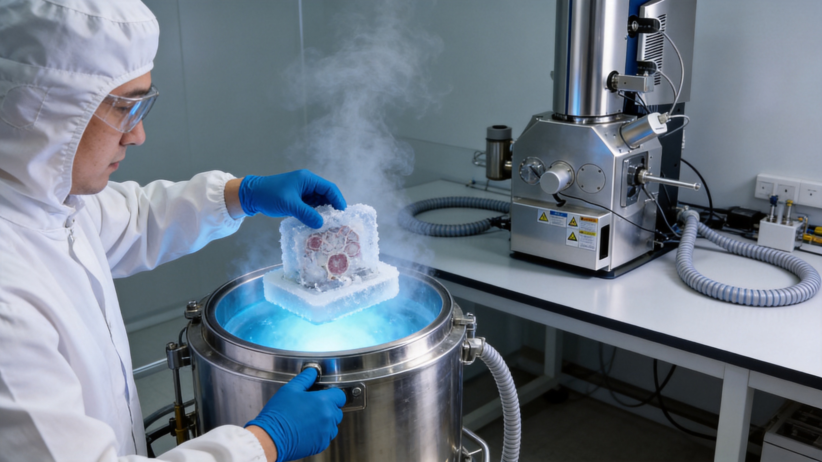
The LSCT series is intended for laboratories that need to maintain frozen samples from preparation through observation. Its central purpose is to create a controlled link between cryogenic sample preparation and electron microscopy. This is especially important when a specimen contains water, volatile components, soft phases, or structures that are easily altered by warming.
In conventional handling, every transfer step can become a source of risk. A sample may encounter ambient humidity, airborne particles, fluctuating temperatures, or an uncontrolled transition between vacuum and atmospheric conditions. These exposures can lead to frost formation, contamination films, ice crystallization, or structural changes. Cryogenic and vacuum transfer reduces these risks by making sample movement a controlled part of the preparation process.
The systems are suitable for workflows in which the specimen must remain frozen during transfer, storage, preparation, or in-situ examination. Their application may include cryo-electron tomography, frozen-hydrated biological specimens, soft matter, emulsions, polymers, pharmaceuticals, and water-containing energy materials.
Maintaining the specimen at low temperature can provide several practical advantages. It helps preserve morphology, reduces molecular mobility, limits evaporation of volatile components, and protects delicate interfaces. It also supports more consistent comparison between samples because the thermal history is better controlled. For research teams working with rare or irreplaceable specimens, this control can significantly reduce the risk of preparing a sample that no longer represents its original state.
Another important advantage is workflow continuity. Cryogenic transfer is not simply a transport function; it is part of the chain of evidence connecting the original specimen to the final image. The less interruption between preparation and observation, the greater the confidence that observed features are intrinsic rather than preparation-induced.
IMP120 Multi-Beam Ion Milling Technology
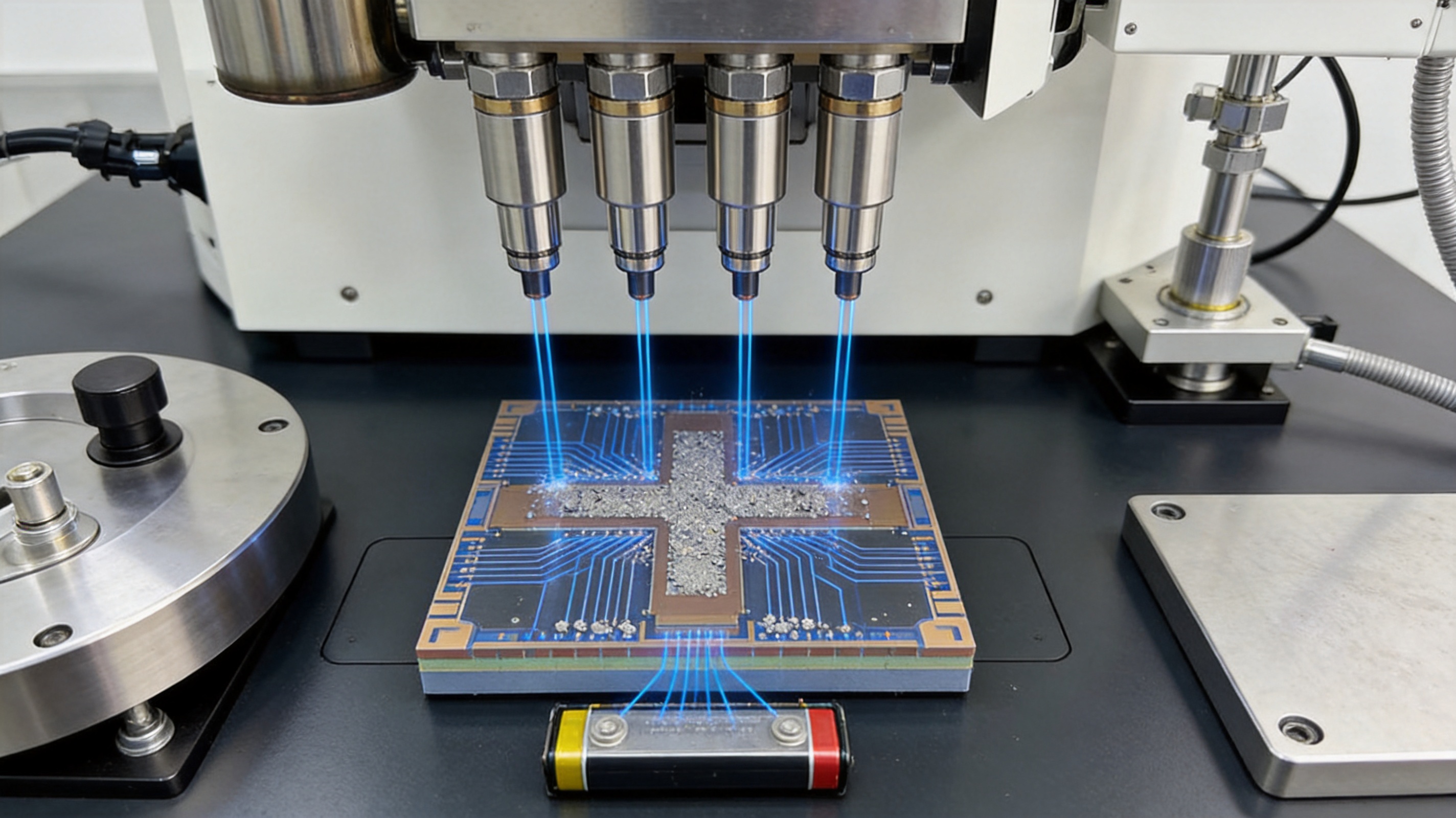
The IMP120 multi-beam ion milling system is designed for precision removal and refinement of material. Ion milling uses accelerated ions to sputter material from a selected region. Because the process is non-contact, it can avoid some of the mechanical forces associated with saw cutting, grinding, and conventional polishing.
Controlled ion milling is valuable when the surface or cross-section must be exceptionally flat, clean, and free from substantial mechanical deformation. It can be used for thinning, polishing, cross-section preparation, cutting, and local surface refinement. The exact process conditions can be selected according to sample composition, thickness, hardness, conductivity, sensitivity, and the desired analytical method.
The multi-beam design supports process flexibility. Different beam arrangements and operating conditions can be used for rough material removal, intermediate preparation, and final polishing. A higher-energy process may be appropriate for efficient removal of bulk material, while a lower-energy finishing process can reduce the depth of the damaged layer and improve the quality of the final observation surface.
This staged approach is particularly useful for advanced materials. Battery electrodes may contain particles, binders, pores, current collectors, and electrolyte residues with very different mechanical properties. Semiconductor devices may combine silicon, metals, dielectrics, polymers, and low-k materials. A single aggressive preparation step can create uneven removal or damage at interfaces. Controlled multi-stage milling allows the operator to balance speed and surface quality.
For transmission electron microscopy, thinning must be performed with careful control of thickness and local geometry. For scanning electron microscopy, the objective may be a broad, flat cross-section with minimal topographic artifacts. For energy-dispersive spectroscopy or other surface-sensitive analysis, contamination and redeposition must also be considered. The IMP120 system supports these varied goals by providing a precision platform for parameter-controlled ion processing.
HSC Series Etching, Coating, and Milling Systems
The HSC series complements cryogenic transfer and multi-beam milling with additional ion-beam preparation functions. Etching can selectively remove material or reveal surface features. Milling can expose internal structures or refine a cross-section. Coating can improve conductivity and reduce charging during electron-beam observation.
Charging is a common problem when non-conductive or poorly conductive specimens are examined in a scanning electron microscope. Excess charge can distort the image, reduce signal stability, create bright regions, or interfere with elemental analysis. A controlled conductive coating can improve beam interaction and make imaging more stable. The appropriate coating material and thickness depend on the sample, the microscope, and whether subsequent chemical or structural analysis will be performed.
HSC systems may also be used for cryogenic treatment of samples that are sensitive to warming. Low-temperature ion processing can help stabilize volatile, hydrated, or soft materials while a surface is prepared. This is valuable when the research objective includes examining a frozen phase, a preserved interface, or a structure that cannot survive ordinary preparation conditions.
By combining etching, coating, and milling in a coordinated preparation environment, the HSC series can reduce the need for repeated manual handling. Fewer transfers can mean lower contamination risk, improved repeatability, and a more efficient laboratory workflow. This is especially important in high-throughput research, contract analysis, and industrial laboratories where consistent preparation quality directly affects productivity.
Major Advantages Over Conventional and Fragmented Approaches
Preservation of Native or Near-Native Structure
The most important advantage of a cryogenic preparation workflow is improved preservation of structure. Biological samples, soft materials, and hydrated interfaces can be examined with less reliance on dehydration or room-temperature exposure. This can help researchers distinguish genuine morphology from artifacts created by drying, collapse, crystallization, or thermal movement.
Reduced Transfer-Related Contamination
Every open transfer creates an opportunity for contamination. Dust, moisture, hydrocarbons, and atmospheric residues can deposit on a specimen and interfere with imaging or spectroscopy. A controlled cryogenic and vacuum transfer route helps protect prepared surfaces between instruments. This is particularly valuable for high-resolution work, where a thin contamination layer can obscure nanoscale details.
Improved Surface Flatness
Ion-beam polishing can produce a smoother and more uniform surface than many mechanical methods. A flatter surface improves imaging consistency, assists interpretation of cross-sectional features, and supports more reliable dimensional measurements. It also helps reduce shadows and topographic interference in surface-sensitive analytical modes.
Lower Mechanical Damage
Mechanical preparation can produce cracks, smearing, pull-out, embedded abrasives, and deformation layers. Ion-beam treatment removes material without direct mechanical contact. Although ion processing must still be carefully controlled to limit implantation, redeposition, heating, and amorphization, it offers a useful route for reducing mechanical artifacts in sensitive specimens.
Greater Material Compatibility
The combined platform is suitable for both soft and hard specimens. Cryogenic transfer addresses samples that require low-temperature preservation, while ion milling and etching support hard materials and multilayer structures. This versatility allows one laboratory to support biological, materials, semiconductor, energy, and pharmaceutical projects without relying on unrelated preparation systems for every application.
More Consistent Reproducibility
Reproducibility depends on controlling the preparation history. Operators can establish process recipes based on temperature, vacuum conditions, beam energy, incidence angle, milling duration, coating conditions, and transfer sequence. A defined procedure is easier to repeat, compare, audit, and optimize than an informal series of manual steps.
Better Integration with Advanced Microscopy
The platform is designed around the needs of modern electron microscopy rather than around a single preparation method. It can support SEM, TEM, cryo-EM, FIB-related workflows, and complementary analytical techniques. This compatibility makes it suitable for laboratories that need to move between imaging modes or study the same sample using multiple methods.
Applications Across Scientific and Industrial Fields
Life Sciences and Cryo-Electron Tomography
In life sciences, the platform supports preparation of cells, tissues, viruses, organelles, and other hydrated specimens. Cryogenic handling helps preserve ultrastructure and spatial relationships. This is useful for cryo-electron tomography, cellular architecture studies, membrane research, pathogen analysis, and synaptic investigations.
For biological research, preparation quality can affect conclusions about organelle shape, membrane continuity, protein complexes, and intracellular organization. A specimen that has been distorted during dehydration may lead to an incorrect interpretation of biological function. Maintaining low-temperature conditions helps researchers work closer to the native state and improves confidence in structural observations.
Materials Science
Materials researchers often need to examine interfaces, defects, grain boundaries, precipitates, pores, and phase distributions. The IMP120 and HSC systems can support cross-section preparation and surface refinement for metals, ceramics, composites, polymers, and advanced functional materials.
For polymer composites, ion processing can help expose the relationship between matrix and reinforcement. For metals, it can assist with analysis of grain boundaries, inclusions, and precipitates. For ceramics, it can reveal pores and interfaces with reduced mechanical smearing. When a material contains volatile or soft phases, cryogenic preparation can add an additional layer of protection.
Semiconductors and Microelectronics
Modern semiconductor devices contain increasingly complex structures and extremely small features. Failure analysis may require a clean cross-section through a contact, transistor, interconnect, dielectric layer, or advanced package interface. Mechanical preparation can damage fragile structures or obscure the location of a defect.
Controlled ion milling and etching can help expose selected regions for SEM or TEM analysis. The ability to combine local material removal with careful surface finishing is valuable for FinFET structures, advanced packaging, multilayer interconnects, memory devices, and process-development investigations. Conductive coating and contamination control can further improve imaging stability.
Energy Materials
Battery and fuel-cell research increasingly focuses on interfaces that change during cycling, heating, charging, and discharging. Examples include solid-electrolyte interphases, electrode particles, current collectors, catalyst layers, and solid-state electrolyte contacts.
These components may be chemically reactive, mechanically fragile, or sensitive to air and moisture. Cryogenic transfer can help preserve selected states, while ion milling can create controlled cross-sections for examining internal architecture. The resulting information can support studies of degradation, crack formation, porosity, interfacial contact, and elemental distribution.
Pharmaceuticals and Nanomedicine
Liposomes, nanoparticles, emulsions, drug carriers, and biological formulations can be difficult to observe because they are soft, hydrated, and beam-sensitive. Cryogenic preparation helps stabilize their morphology. Electron microscopy can then be used to study size distribution, shape, aggregation, encapsulation-related structures, and interaction with biological environments.
Geology and Mineralogy
Geological specimens often contain hard and heterogeneous phases. Ion-beam processing can assist in preparing clean cross-sections through mineral inclusions, shale pores, interfaces, and microstructural features. A controlled surface is important when imaging is combined with elemental analysis or when the research focuses on nanoscale pore networks.
Third-Party Testing and Analytical Services
Testing laboratories and analytical service providers require dependable preparation methods because they serve customers with widely different sample types. A versatile platform allows these laboratories to process frozen biological specimens, battery materials, semiconductor components, polymers, and industrial materials using controlled procedures. Consistent preparation can improve turnaround time, reduce repeat work, and strengthen the credibility of analytical reports.
Integrated Workflow Examples
Example One: Cryogenic Biological Preparation
A biological specimen may first be rapidly frozen to immobilize water and cellular structures. It can then be introduced into a cryogenic preparation environment, where surface treatment or fracture is performed under suitable conditions. The LSCT system transfers the frozen sample through a controlled route to the electron microscope. If coating is required, an HSC system can provide an appropriate conductive or functional layer while preserving the low-temperature state.
The objective is to minimize warming, ice formation, contamination, and morphological collapse. This workflow is appropriate for cryo-ET, pathogen studies, cellular ultrastructure, and other investigations in which native or near-native organization is essential.
Example Two: Cryogenic Ion Polishing of an Energy Material
A battery electrode or solid-state electrolyte may be prepared and stabilized at low temperature. The IMP120 system can then be used for controlled ion polishing or cross-section refinement. After processing, the LSCT system can support cryogenic transfer to the microscope, limiting environmental exposure.
This approach can help preserve interfaces and volatile components that might be altered during room-temperature preparation. Researchers may then examine particle contacts, interphase layers, cracks, pores, and reaction products with improved confidence.
Example Three: Semiconductor Cross-Section Analysis
A selected region of a semiconductor device can be exposed through controlled cutting and ion milling. Rough removal may be followed by a lower-energy finishing step to improve the final surface. If charging affects the observation, the HSC system can be used for a suitable coating process. The completed cross-section can then be examined by SEM, TEM, or complementary analytical methods.
This workflow is designed to reduce mechanical deformation and improve the visibility of small features. It can support failure analysis, process development, quality verification, and advanced packaging inspection.
Example Four: Polymer and Composite Interface Study
A polymer composite can be cooled to reduce deformation during preparation. Ion-beam milling can expose a cross-section through the reinforcement and matrix. Subsequent coating can improve conductivity for SEM imaging. The prepared surface may then be analyzed for bonding quality, voids, particle dispersion, delamination, and interface morphology.
Manufacturing and Engineering Strengths
JIANGSU BAISHENG INDUSTRIAL CO., LTD. approaches the product family through a combination of engineering development, precision manufacturing, and international supply experience. The company was founded in 2010 and has developed from an electronic testing research and development studio into a technology-oriented enterprise providing laboratory equipment and safety testing instruments.
The company’s history supports a practical understanding of laboratory equipment development. In 2013, its predecessor focused on electronic testing research and development. In 2016, the organization moved toward independent production and launched laboratory equipment with its own technical development capabilities. In 2019, it adopted a technology-plus-trade strategy, increasing research investment, adding technical personnel, and expanding its overseas market activities. By 2022, the company had further strengthened its quality management system and technical specification processes.
This background is relevant to the Electron Microscopy Sample Preparation Platform because advanced preparation equipment requires more than basic fabrication. It requires the coordination of mechanical structures, vacuum-compatible components, thermal management, ion-beam control, electrical systems, user interfaces, safety functions, and process repeatability. Experience in electronic testing and laboratory equipment provides a foundation for integrating these elements into usable instruments.
The company emphasizes precision in craftsmanship and long-term innovation. In practice, this means that manufacturing quality should be considered across the complete product life cycle, from engineering design and component selection to assembly, inspection, commissioning, packaging, and technical support.
Design and Process Engineering
Engineering begins with the sample and the intended microscopy workflow. A useful system must accommodate different specimen dimensions, holders, preparation sequences, temperature conditions, and vacuum requirements. Modular design principles can make it easier to adapt the equipment to different applications while maintaining stable core functions.
Process engineering also includes the selection of materials and components that can perform reliably under vacuum, low temperature, electrical load, and ion-beam exposure. Mechanical stability is important because vibration or movement can affect beam alignment and surface uniformity. Thermal stability is important because uncontrolled temperature change may compromise cryogenic specimens or alter milling behavior.
Precision Assembly and Alignment
Ion-beam systems depend on accurate alignment between the sample, beam, milling area, and observation position. Transfer systems depend on reliable movement, sealing, holder compatibility, and environmental control. Assembly quality therefore has a direct effect on practical performance.
Precision assembly includes dimensional verification, fit checks, cable and connection inspection, vacuum-seal verification, motion testing, and functional commissioning. Proper alignment and repeatable positioning help operators reproduce preparation conditions from one sample to the next.
Quality Control and Verification
Quality control for laboratory equipment should extend beyond visual inspection. Functional testing may include vacuum performance, thermal response, movement accuracy, beam stability, coating operation, safety interlocks, and system communication. Inspection records can help identify deviations and provide traceability during delivery and service.
For customers, this approach reduces the risk associated with installing a high-value instrument in a research or production environment. It also creates a foundation for application-specific commissioning and method development.
Customization and Application Support
Electron microscopy laboratories rarely have identical requirements. One customer may prioritize cryogenic transfer, while another may need large-area cross-sectioning, low-energy polishing, conductive coating, or special sample holders. A manufacturer with an engineering-oriented R&D team can discuss the actual workflow and configure the equipment accordingly.
Customization may involve holder design, chamber layout, transfer interfaces, process parameters, accessory selection, software functions, or integration with existing microscopes. The goal is not customization for its own sake, but a better match between the equipment and the samples, instruments, and working practices of the laboratory.
Operational Benefits for Research and Industry
The integrated platform can improve laboratory efficiency by reducing unnecessary manual handling. When sample preparation is performed with separate, disconnected tools, the operator may need to expose the specimen repeatedly to air, change holders several times, or move it between incompatible workstations. Each additional step can increase preparation time and introduce variability.
A coordinated platform supports more structured workflows. Operators can establish standard operating procedures for biological specimens, battery materials, semiconductor cross-sections, polymers, and other sample classes. These procedures can include sample mounting, thermal conditioning, rough milling, finishing, coating, transfer, and observation.
Standardization also improves communication between researchers. When the preparation history is documented, results can be compared more meaningfully. Researchers can distinguish differences in sample behavior from differences caused by operator technique. In industrial environments, documented preparation conditions can support quality assurance, root-cause analysis, and customer reporting.
The equipment can also contribute to resource efficiency. Better preparation reduces the number of failed specimens, repeated microscope sessions, and discarded samples. For rare biological material or expensive advanced materials, this benefit can be especially significant.
Recommended Preparation Considerations
Although the platform is designed for flexibility, successful results still depend on appropriate process development. Operators should begin by defining the analytical objective. The requirements for a broad SEM cross-section are different from those for a TEM lamella, cryo-ET specimen, elemental analysis surface, or interface study.
Sample composition should be considered before selecting beam conditions. Materials with different sputtering rates may develop uneven surfaces if processed too aggressively. Multilayer devices may require staged milling. Soft or volatile materials may require low-temperature handling. Non-conductive samples may need coating, but coating thickness must be compatible with the intended observation and chemical analysis.
Temperature control should be treated as a process variable rather than a secondary convenience. The sample’s freezing history, transfer duration, exposure conditions, and observation temperature can all influence the final result. For cryogenic work, operators should also consider frost prevention, holder cleanliness, vacuum readiness, and the time required to move the sample between stages.
Beam energy and incidence angle should be optimized for the sample and the desired finish. Higher-energy processing can increase material removal rates, while lower-energy finishing can help improve surface quality and reduce near-surface damage. A staged procedure is often more effective than using one condition for the entire operation.
Finally, the preparation record should be retained. Recording sample identity, temperature, vacuum condition, beam parameters, processing duration, coating details, and transfer sequence makes troubleshooting easier and supports reliable comparison across experiments.
Competitive Positioning
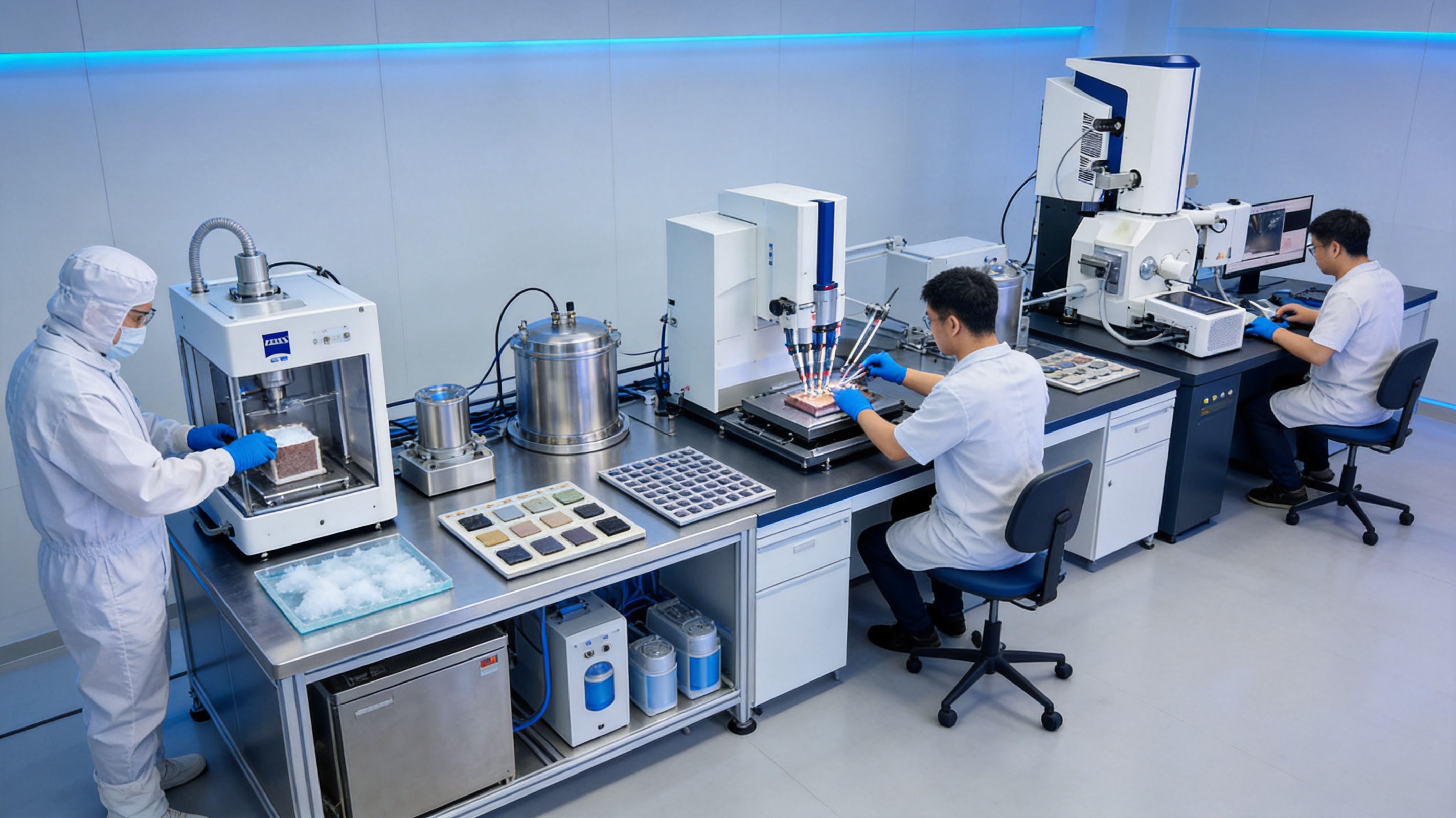
The platform’s competitive strength comes from combining functions that are often purchased and operated separately. A laboratory may otherwise need one system for cryogenic transfer, another for ion milling, another for coating, and additional accessories for sample movement. Separate systems can be effective, but they may create compatibility issues and increase the number of handling steps.
An integrated equipment family offers a more coherent technical route. Cryogenic transfer supports preservation. Ion milling supports surface and cross-section quality. Etching and coating support observation readiness. Together, these capabilities address the most common sources of preparation error: warming, contamination, deformation, roughness, charging, and inconsistent processing.
Another differentiating factor is application breadth. Some preparation systems are optimized for either biological cryo-work or hard-material cross-sections. This platform is intended to cover both areas through complementary configurations. That makes it attractive to shared microscopy facilities, universities, national laboratories, industrial R&D centers, and testing organizations that serve multiple scientific disciplines.
The company’s combination of R&D capability and international trade experience is also valuable for customers outside China. Technical equipment must be accompanied by clear communication, documentation, installation coordination, spare-parts planning, and after-sales support. A supplier that understands both engineering requirements and international project execution can make adoption easier for overseas laboratories.
Compared with low-cost, single-function equipment, the platform is positioned around process control and sample fidelity. Its value is measured not only by the initial purchase price, but also by the quality of images obtained, the reduction of repeated preparation, the protection of valuable samples, and the ability to expand laboratory capabilities over time.
Customer Groups and Procurement Value
Typical users include universities, research institutes, electron microscopy centers, national laboratories, hospitals, biomedical research centers, semiconductor manufacturers, new-energy companies, pharmaceutical enterprises, advanced-materials producers, and third-party testing organizations.
For universities and research institutes, the platform provides a flexible foundation for multidisciplinary projects. A shared facility can support biological cryo-analysis in one project and battery or semiconductor analysis in another. For hospitals and biomedical centers, the cryogenic workflow can assist with pathogen, cellular, and pharmaceutical research. For industrial customers, precision cross-sectioning and surface treatment can support process control, failure analysis, and product development.
Procurement teams should evaluate the equipment according to the complete workflow rather than a single specification. Important considerations include sample types, required temperature range, vacuum needs, beam-processing capability, coating requirements, compatibility with existing microscopes, available laboratory space, operator training, maintenance support, and future expansion plans.
Purchasing consideration |
Why it matters |
Relevant platform capability |
Sample preservation |
Protects hydrated, volatile, and beam-sensitive specimens |
LSCT cryogenic and vacuum transfer; cryogenic HSC processing |
Cross-section quality |
Improves visibility of interfaces, defects, and internal structures |
IMP120 and HSC ion milling and etching |
Surface conductivity |
Reduces charging during electron-beam imaging |
HSC coating functions |
Workflow integration |
Reduces handling and transfer-related variation |
Coordinated transfer and preparation ecosystem |
Future flexibility |
Supports changing research and production requirements |
Complementary equipment series and customization support |
Implementation and Training
Successful installation begins with a review of the laboratory environment. The customer and supplier should confirm floor space, power supply, ventilation, cooling requirements, vacuum infrastructure, gas arrangements where applicable, microscope interfaces, sample-holder standards, and operator access.
Training should cover both equipment operation and preparation methodology. Operators need to understand how sample composition affects beam processing, how cryogenic conditions influence transfer, how coatings affect imaging, and how to identify signs of contamination or preparation damage. Training is most effective when it includes representative customer samples rather than only generic demonstrations.
Application development may begin with a small set of standard specimens. The team can compare different beam energies, milling durations, angles, coating conditions, and transfer procedures. Once the desired surface or preservation quality has been achieved, the process can be documented as a laboratory recipe.
Preventive maintenance should be incorporated into the operating plan. Cleaning, consumable replacement, vacuum checks, holder inspection, alignment verification, and software updates help maintain stable performance. A regular maintenance schedule is particularly important for shared facilities with high instrument utilization.
Future Development of Electron Microscopy Preparation
Electron microscopy is moving toward higher resolution, more complex in-situ experiments, multimodal characterization, and increasingly automated workflows. As microscope performance improves, preparation artifacts become more visible and more consequential. The demand for cleaner surfaces, thinner specimens, better cryogenic preservation, and more repeatable processes is therefore expected to continue.
Digital process records may become increasingly important. Linking preparation parameters with microscope images and analytical results can support machine-assisted optimization, statistical process control, and more reliable comparison between laboratories. Automation may also reduce operator-to-operator variation in transfer, milling, coating, and inspection.
At the same time, new materials will create new preparation challenges. Solid-state batteries, advanced semiconductor packages, quantum materials, biological therapeutics, and complex polymer systems may combine phases with very different thermal, electrical, and mechanical properties. Flexible preparation platforms will be better positioned to respond to these requirements than narrowly specialized systems.
The long-term importance of the platform is therefore broader than any single instrument. It provides a technical foundation for improving the connection between sample preparation and microscopy interpretation. By preserving the sample more effectively and preparing surfaces more precisely, it helps researchers obtain data that are more representative, repeatable, and useful.
Questions and Answers
What is the main purpose of the Electron Microscopy Sample Preparation Platform?
The main purpose is to prepare high-quality specimens for electron microscopy while preserving sample state, reducing contamination, and improving surface or cross-section quality. It combines cryogenic transfer with ion-beam milling, etching, polishing, thinning, cutting, and coating functions.
Which instruments are included in the platform?
The platform includes LSCT series cryogenic transfer systems, the IMP120 multi-beam ion milling system, and HSC series ion beam etching, coating, and milling systems. The instruments can be used independently or as parts of a coordinated workflow.
Why is cryogenic transfer important?
Cryogenic transfer helps maintain frozen conditions and limits exposure to moisture, contaminants, and warming. It is especially important for hydrated biological specimens, soft materials, volatile samples, and interfaces that may change at room temperature.
Can the platform be used for hard materials?
Yes. The IMP120 and HSC systems are suitable for metals, ceramics, semiconductors, battery materials, minerals, and other hard or heterogeneous specimens. They can support cross-section preparation, polishing, thinning, cutting, and surface refinement.
Is the equipment suitable for biological research?
Yes. LSCT cryogenic transfer systems and cryogenic preparation functions can support cells, tissues, viruses, organelles, and other hydrated or beam-sensitive samples. Typical applications include cryo-electron tomography, cellular ultrastructure, pathogen analysis, and pharmaceutical nanostructure research.
How does ion milling compare with mechanical polishing?
Ion milling is a non-contact material-removal process. When properly controlled, it can reduce mechanical scratches, smearing, deformation, and abrasive contamination. Mechanical preparation may still be useful for bulk removal, but ion milling is valuable for precision finishing and sensitive cross-sections.
What is the purpose of coating a sample?
Coating can improve electrical conductivity and reduce charging during SEM observation. It may also enhance signal stability and image quality. Coating conditions should be selected carefully when the sample will undergo elemental, surface-sensitive, or high-resolution analysis.
Can the systems be customized?
Configuration and workflow requirements can be discussed according to sample type, microscope model, holder design, temperature needs, beam-processing objectives, coating requirements, and laboratory layout. Customization should be based on the customer’s analytical process and future application plans.
Who are the typical customers?
Typical customers include universities, research institutes, national laboratories, hospitals, microscopy centers, semiconductor companies, new-energy manufacturers, pharmaceutical companies, advanced-materials producers, and third-party testing organizations.
What should customers prepare before purchasing?
Customers should identify their sample types, target microscopy methods, required cross-section or surface quality, cryogenic needs, coating requirements, available infrastructure, expected sample volume, and operator skill level. This information helps determine the most appropriate system combination and workflow.
Conclusion
The Electron Microscopy Sample Preparation Platform addresses a central challenge in modern microscopy: obtaining images and analytical data that faithfully represent the original specimen. LSCT cryogenic transfer systems help protect frozen and hydrated samples. The IMP120 multi-beam ion milling system provides controlled removal, thinning, polishing, and cross-section preparation. HSC series systems add ion-beam etching, coating, milling, and cryogenic treatment capabilities.
Used together, these technologies create a complete preparation ecosystem for life sciences, materials science, semiconductors, energy materials, pharmaceuticals, geology, and industrial analysis. Their principal advantages include improved sample preservation, reduced contamination, lower mechanical damage, better surface flatness, enhanced reproducibility, and greater workflow flexibility.
JIANGSU BAISHENG INDUSTRIAL CO., LTD. supports this product direction with a technology-driven development model, an R&D background in electronic testing and laboratory equipment, independent production experience, quality management development, and international trade capability. Its emphasis on precision craftsmanship, long-term innovation, and customized solutions positions the company to serve laboratories seeking dependable preparation equipment for demanding electron microscopy applications.
As microscopy continues toward higher resolution and more complex materials, sample preparation will remain a decisive factor in analytical quality. A controlled platform that combines cryogenic preservation with precision ion-beam processing gives researchers and industrial users a stronger foundation for accurate, repeatable, and high-value microscopy results.
References
1. Goldstein, J., Newbury, D., Joy, D., Lyman, C., Echlin, P., Lifshin, E., Sawyer, L., and Michael, J. Scanning Electron Microscopy and X-Ray Microanalysis. Springer.
2. Williams, D. B., and Carter, C. B. Transmission Electron Microscopy: A Textbook for Materials Science. Springer.
3. Frank, J. Cryo-Electron Microscopy and Tomography: Principles and Applications. Academic and scientific reference literature.
4. Reimer, L. Scanning Electron Microscopy: Physics of Image Formation and Microanalysis. Springer.
5. Egerton, R. F. Physical Principles of Electron Microscopy: An Introduction to TEM, SEM, and AEM. Springer.
6. International Organization for Standardization. General guidance on laboratory competence, quality management, and measurement traceability.
7. Relevant manufacturer operating procedures, application notes, and technical documentation for cryogenic transfer, ion milling, ion etching, and conductive coating systems.
8. International guidance and laboratory best practices for cryo-electron microscopy sample handling, contamination control, vacuum transfer, and beam-sensitive specimen preparation.
Product: Electron Microscopy Sample Preparation Platform

PREV:Multi-Beam Ion Beam Processing Systems for Precision Surface Engineering
NEXT:Cryogenic Environmental Chambers for Reliable Low-Temperature Sample Preparation and Analysis
NEXT:Cryogenic Environmental Chambers for Reliable Low-Temperature Sample Preparation and Analysis
Interested in cooperation or have questions?
Featured Products
Related products
Quick Link
Contact Information
- Tel: +86-13861043568
- E-mail: 1840784112@qq.com
- Add: Tianting District, Changzhou City, Jiangsu Province, China, No. 6 Hecircuit
Scan Code
Copyright © JIANGSU BAISHENG INDUSTRIAL CO., LTD.
All Rights Reserved. Privacy Custom Precision Laboratory Testing Equipment Manufacturers
