 English
 Español
NEWS
NEWS
NEWS
NEWS
NEWS
NEWS
NEWS
NEWS
NEWS
NEWS
NEWS
NEWS
NEWS
NEWS
NEWS
NEWS

Luo Qinyue — Senior After-Sales Service Engineer
Home / Author / Luo Qinyue — Senior After-Sales Service Engineer / Advanced Electron Microscopy Sample Preparation Platforms for High-Fidelity Analysis
Advanced Electron Microscopy Sample Preparation Platforms for High-Fidelity Analysis
Content
- 1 The Importance of Sample Preparation in Electron Microscopy
- 2 Integrated Platform Architecture
- 3 LSCT-Series Cryogenic Transfer Systems
- 4 IMP120 Multi-Beam Ion Milling Technology
- 5 HSC-Series Ion Beam Etching, Coating, and Milling Systems
- 6 Advantages Over Conventional and Disconnected Preparation Workflows
- 7 Advanced Manufacturing and Engineering Strengths
- 8 Application in Life Sciences and Cryo-Electron Microscopy
- 9 Application in Materials Science
- 10 Application in Semiconductors and Microelectronics
- 11 Application in Geology, Pharmaceuticals, and Energy Research
- 12 Recommended Workflow for High-Fidelity Sample Preparation
- 13 Operational and Commercial Benefits
- 14 Quality, Reliability, and Long-Term Development
- 15 Choosing the Right Configuration
- 16 Why This Platform Is a Strong Competitive Choice
- 17 Questions and Answers
- 17.1 What is an electron microscopy sample preparation platform?
- 17.2 Which electron microscopes can use the platform?
- 17.3 Why is cryogenic transfer important?
- 17.4 Can hard materials be processed with the same platform?
- 17.5 What is the benefit of multi-beam ion milling?
- 17.6 Can the platform process biological samples?
- 17.7 Is coating always necessary?
- 17.8 How does the platform improve reproducibility?
- 17.9 Can the equipment be customized?
- 17.10 Who are the primary users?
- 17.11 What should customers consider before purchasing?
- 18 Conclusion
- 19 References
- 20 Product: Electron Microscopy Sample Preparation Platform
Modern electron microscopy can reveal structures and interfaces at scales that are inaccessible to optical methods. However, the quality of an electron microscopy result depends on much more than the performance of the microscope itself. Sample preparation, environmental control, transfer integrity, surface condition, and protection against contamination or radiation damage all influence the reliability of the final image and analytical data.
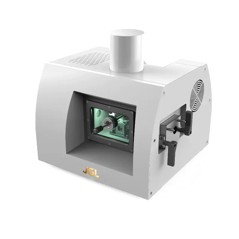
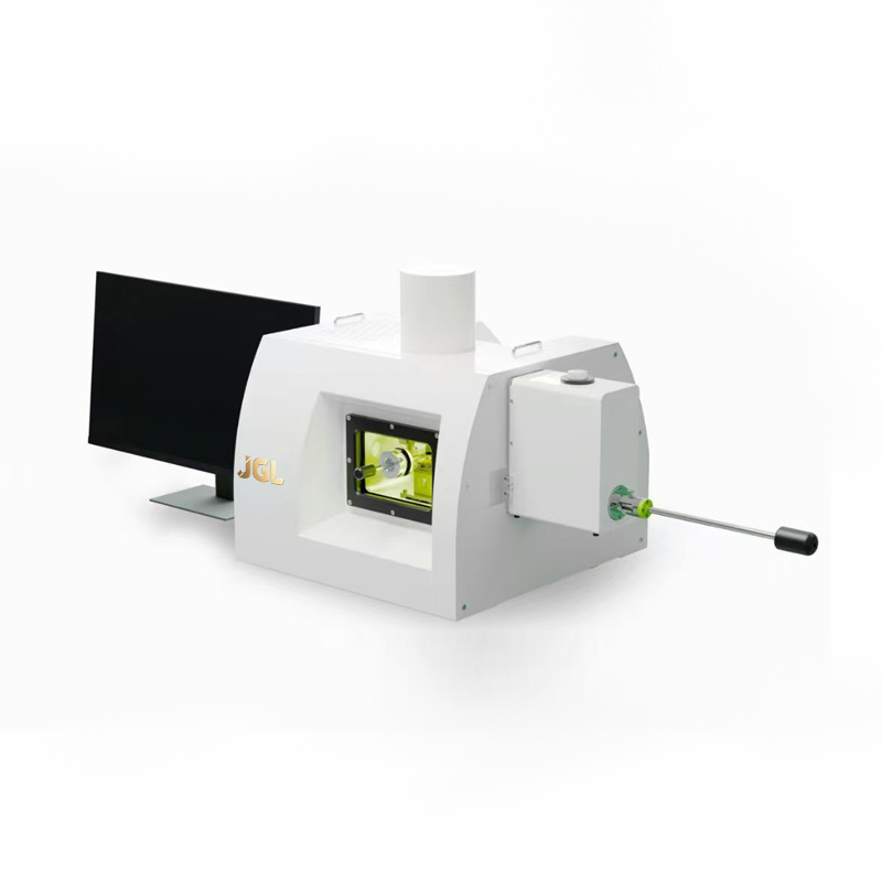
The Electron Microscopy Sample Preparation Platform is an integrated family of auxiliary systems developed for demanding sample preparation, cryogenic transfer, ion beam processing, coating, polishing, thinning, and in-situ or cryogenic observation. The platform combines LSCT-series cryogenic transfer systems, IMP120 multi-beam ion milling technology, and HSC-series ion beam etching, coating, and milling systems. Together, these instruments support a complete workflow for preparing high-quality samples for scanning electron microscopy, transmission electron microscopy, focused ion beam workflows, cryo-electron microscopy, and related analytical methods.
Designed for laboratories, research institutes, industrial R&D departments, analytical service providers, and advanced manufacturing organizations, the platform addresses a broad range of sample types. These include biological specimens, hydrated materials, polymers, soft matter, metals, ceramics, semiconductor structures, battery materials, geological samples, pharmaceutical formulations, and nanostructured systems.
Its central value is the ability to preserve sample fidelity while delivering the surface quality, cross-sectional accuracy, and environmental stability required by advanced electron microscopy. Instead of treating cryogenic transfer, ion milling, coating, and sample preparation as isolated operations, the platform connects them into a coordinated technical ecosystem.
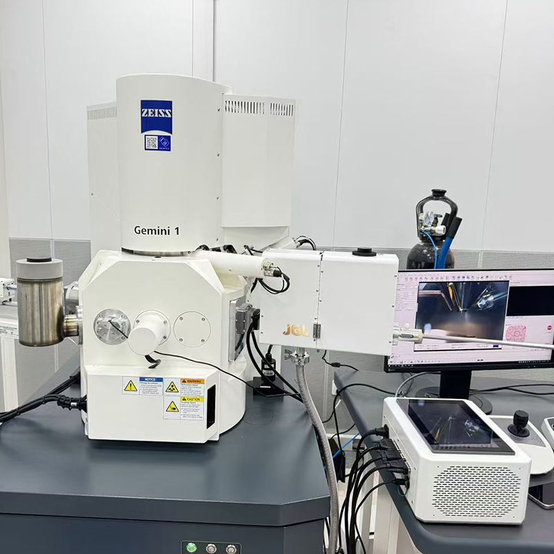
Electron Microscopy Sample Preparation Platform
The Importance of Sample Preparation in Electron Microscopy
Electron microscopes operate with highly focused electron beams and are capable of producing detailed structural, chemical, and topographical information. Nevertheless, electron microscopy cannot correct a poorly prepared specimen. Dehydration, contamination, charging, mechanical deformation, curtaining, ion-beam damage, redeposition, and ice crystallization can all create artifacts that obscure the true structure of a sample.
Biological specimens are especially sensitive. Cells, tissues, viruses, membrane systems, and protein assemblies contain significant amounts of water and may collapse or undergo chemical changes during conventional drying. A rapid cryogenic fixation process can immobilize water and biological structures, preserving a state closer to the native hydrated condition. That benefit is only maintained if the frozen specimen remains cold and protected throughout transfer and observation.
Soft materials and polymers present a different set of challenges. Their mechanical properties may change significantly when temperature or pressure changes. Vacuum exposure can cause outgassing, shrinkage, or loss of volatile components. Electron-beam exposure may also produce local heating, cross-linking, decomposition, or charging. Controlled low-temperature preparation can reduce these effects and improve the reliability of morphological observations.
Hard materials require precision rather than hydration preservation. Battery electrodes, semiconductor layers, metallic interfaces, ceramic composites, and geological specimens often need a flat cross-section or a thin electron-transparent region. Mechanical preparation alone can introduce scratches, stress, smearing, or fractured edges. Ion beam processing provides a controllable method for removing material and refining surfaces with minimal mechanical contact.
For all of these applications, preparation is not merely a preliminary laboratory task. It is a critical part of the measurement chain. A platform that stabilizes the sample from preparation through transfer and imaging can therefore improve reproducibility, reduce rework, and increase confidence in analytical conclusions.
Integrated Platform Architecture
The platform is organized around three complementary technology groups. The LSCT series supports cryogenic and vacuum transfer. The IMP120 multi-beam ion milling system provides precision processing under ambient or cryogenic conditions. The HSC series provides ion beam etching, coating, and milling functions for a broad range of sample preparation requirements.
Each system can serve a specific laboratory need, but the greatest value is achieved when the instruments are used as a coordinated workflow. For example, a frozen biological sample may undergo cryogenic preparation, remain under controlled low-temperature conditions during transfer, and then be introduced into a cryo-electron microscope without exposure to ambient moisture. Similarly, a battery cross-section may be ion polished to reveal an interface, coated for improved conductivity, and transferred for high-resolution imaging and elemental analysis.
This modular architecture allows users to configure the platform according to application requirements. A laboratory focused on life sciences may prioritize cryogenic transfer and contamination control. A semiconductor laboratory may emphasize cross-sectional preparation, precision milling, and interface inspection. A third-party testing center may require a flexible combination of cryogenic and room-temperature capabilities to support many customer sample types.
| Platform component | Primary function | Typical contribution to the EM workflow |
| LSCT series | Cryogenic and vacuum transfer | Preserves frozen samples and minimizes ice growth, contamination, and atmospheric exposure |
| IMP120 | Multi-beam ion milling and polishing | Produces clean, flat, and precisely processed surfaces under ambient or cryogenic conditions |
| HSC series | Ion beam etching, coating, and milling | Supports surface refinement, conductive coating, thinning, and specialized specimen preparation |
| Integrated workflow | Preparation, transfer, and observation support | Improves sample continuity, process control, and analytical reproducibility |
LSCT-Series Cryogenic Transfer Systems
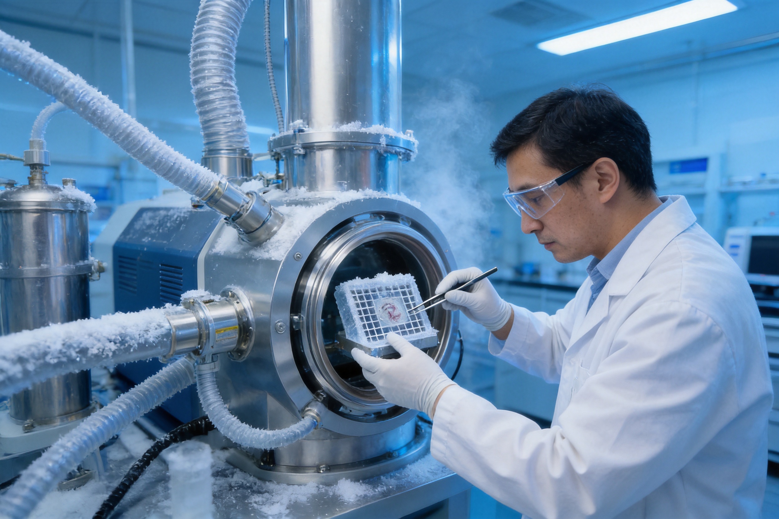
The LSCT series is designed for the controlled movement of frozen or temperature-sensitive specimens between preparation equipment, storage environments, and electron microscopes. Its purpose is to protect the sample from the conditions that can compromise cryogenic integrity, including moisture, contamination, temperature rise, and uncontrolled exposure to the surrounding atmosphere.
In cryo-electron microscopy, the period between sample preparation and observation is often as important as the initial freezing step. A specimen that has been correctly vitrified can still be damaged by warming or by the formation of crystalline ice during transfer. Even a small amount of contamination may interfere with imaging, obscure surface features, or affect downstream analysis.
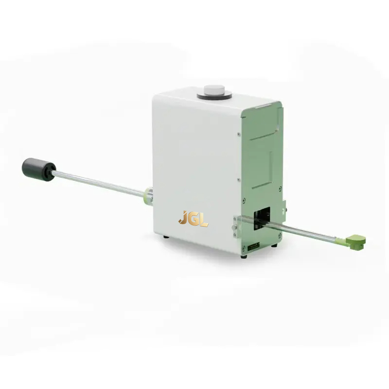
By supporting cryogenic and vacuum transfer, LSCT systems help create a continuous protected path. The exact configuration can be adapted to the requirements of the sample holder, preparation instrument, and electron microscope. Models such as the LSCT100, LSCT102, and LSCT200 represent a product family intended to support different levels of transfer functionality and laboratory integration.
The system concept is particularly valuable for cryo-electron tomography, where the three-dimensional reconstruction of cells, organelles, viruses, and macromolecular assemblies requires the preservation of structural relationships. Any collapse, contamination, or ice artifact can reduce the quality of the reconstructed volume. Maintaining a stable cryogenic state allows the microscope to examine a specimen that more closely represents its prepared biological condition.
LSCT technology is also relevant to non-biological materials. Electrolytes, hydrated polymers, emulsions, soft composites, and water-containing nanomaterials may change rapidly when exposed to vacuum or room temperature. Cryogenic transfer can retain volatile phases and preserve interfaces that would otherwise be lost.
Protection Against Ice Crystallization
Water can exist in different solid states depending on the cooling rate and environmental conditions. In many cryo-electron microscopy applications, vitrification is preferred because it immobilizes water in an amorphous state and reduces the disruption associated with ice crystal formation. Once a sample is vitrified, maintaining the correct temperature is essential.
The LSCT series supports the operational discipline required after freezing. By limiting exposure during transfer, it helps reduce the risk of frost accumulation and crystallization. This is important for biological ultrastructure, where membranes, protein complexes, and cellular compartments may be distorted by crystalline ice.
Vacuum-Compatible Transfer
Vacuum compatibility is another important feature of a cryogenic transfer workflow. Atmospheric transfer can introduce water vapor, airborne particles, and other contaminants. A vacuum-supported path can reduce those risks and create a cleaner interface between preparation equipment and the electron microscope.
For advanced laboratories, vacuum transfer also supports more consistent process documentation. Operators can define transfer sequences, monitor critical conditions, and reduce the variability associated with manual handling. This is especially beneficial when multiple researchers use the same facility or when samples must be prepared according to validated procedures.
IMP120 Multi-Beam Ion Milling Technology
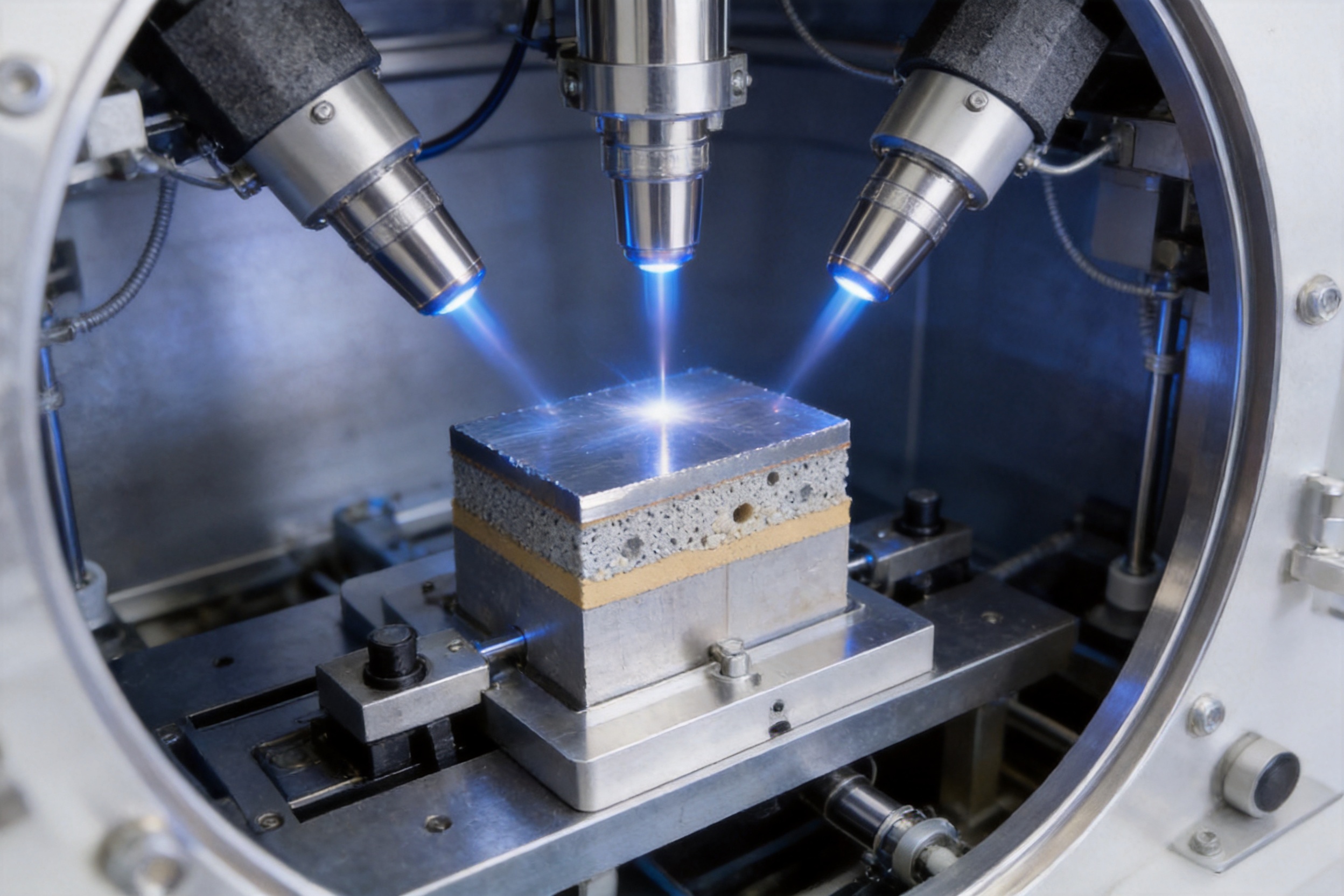
The IMP120 multi-beam ion milling system is intended for high-quality sample surface preparation, thinning, polishing, and cross-section refinement. Ion milling removes material through controlled bombardment, allowing the operator to process samples without applying the mechanical forces associated with conventional grinding or polishing.
Mechanical preparation remains useful for reducing bulk material, but it may leave scratches, deformation layers, embedded abrasives, or smeared phases. These artifacts can be especially problematic in heterogeneous materials, where different phases respond differently to mechanical stress. Ion milling provides a final refinement step that can expose a cleaner and more representative structure.
The multi-beam configuration expands the range of preparation conditions available to the operator. Depending on the sample and the desired result, the process can be adjusted for material removal, surface finishing, local thinning, or preparation under reduced-temperature conditions. This flexibility is important because biological samples, battery materials, semiconductor devices, and metals do not respond identically to ion bombardment.
Ambient and Cryogenic Processing
A major advantage of the IMP120 concept is its ability to support both ambient and cryogenic preparation. Ambient ion milling is suitable for many metals, ceramics, semiconductors, geological specimens, and other stable materials. Cryogenic processing is appropriate for soft, volatile, hydrated, or beam-sensitive specimens.
Low-temperature milling can reduce the risk of thermal alteration during ion bombardment. For polymers and biological materials, cooling may help retain morphology and reduce the mobility of water or volatile components. For battery materials, cryogenic preparation may help preserve electrolyte-related features and unstable interfaces that are difficult to observe after warming.
The combination of ion processing and temperature control gives laboratories more options than a single-condition milling system. It also reduces the need to move sensitive specimens between unrelated preparation environments, which can introduce handling errors and increase the possibility of contamination.
Surface Quality and Cross-Section Accuracy
A good electron microscopy surface should be sufficiently flat, clean, and free from preparation-induced structures. This is particularly important for high-resolution imaging, backscattered electron contrast, energy-dispersive X-ray spectroscopy, electron energy-loss spectroscopy, and automated image analysis.
Ion milling can be used to remove damaged layers and expose a cross-section through an interface or feature of interest. In a battery electrode, for example, the quality of the prepared cross-section can influence the interpretation of particle cracking, binder distribution, pore structure, and solid-electrolyte interphase layers. In semiconductor devices, a controlled cross-section can reveal gate structures, dielectric layers, contacts, voids, and packaging interfaces.
The IMP120 is therefore not limited to producing visually attractive surfaces. Its role is to improve the analytical value of the specimen by reducing preparation artifacts that might otherwise be mistaken for real material features.
HSC-Series Ion Beam Etching, Coating, and Milling Systems
The HSC series extends the platform with functions for ion beam etching, coating, and milling. These processes are important when sample conductivity, surface composition, topographic definition, or localized material removal must be controlled before observation.
Ion beam etching can selectively refine a surface or expose a region beneath the original exterior. Milling can remove material to create a cross-section, cavity, or thin area. Coating can improve electrical conductivity, reduce charging, enhance signal stability, or provide a controlled surface condition for imaging.
These capabilities support both routine and advanced workflows. A laboratory may use coating to prepare nonconductive biological or polymer samples for scanning electron microscopy. It may use milling to inspect a multilayer electronic structure. It may combine etching and coating to improve contrast or analytical stability.
The HSC family is also useful when the sample requires a specialized preparation sequence that cannot be achieved through mechanical methods alone. The ability to integrate several operations within a coordinated platform can reduce transfer steps and help maintain sample orientation.
Conductive Coating for Stable Imaging
Nonconductive specimens can accumulate charge under an electron beam. Charging may cause image drift, brightness fluctuations, distortion, or difficulty in obtaining stable analytical signals. A thin conductive coating can help dissipate charge and improve imaging behavior.
Coating must be selected carefully. Excessive thickness may obscure fine surface features, alter analytical results, or reduce spatial resolution. Insufficient or uneven coating may not solve charging problems. A controlled coating system enables the operator to match the layer and process to the sample type and imaging objective.
For delicate specimens, coating may be performed after cryogenic preparation or under conditions designed to minimize thermal and structural disturbance. For hard materials, it may be used as part of a broader cross-sectional or surface-analysis workflow.
Advantages Over Conventional and Disconnected Preparation Workflows
The platform offers several advantages when compared with workflows based on disconnected instruments or purely mechanical preparation methods. These advantages relate to sample preservation, process continuity, application flexibility, surface quality, and operational efficiency.
1. Better Preservation of Native or Prepared States
For cryogenic and temperature-sensitive specimens, preserving the sample state is often the primary requirement. Conventional room-temperature transfer can result in drying, collapse, contamination, or phase changes. The LSCT series provides a protected route that helps maintain the intended condition from preparation to observation.
This benefit is particularly relevant to biological research, where structural fidelity is directly connected to scientific interpretation. It is also valuable for materials containing volatile electrolytes, solvents, water, or soft phases that are easily altered during ordinary handling.
2. Lower Risk of Mechanical Damage
Mechanical polishing can create deformation and smearing, especially when the specimen contains phases with different hardness or ductility. Ion beam processing removes material without direct mechanical contact and can serve as a final precision step after bulk preparation.
For layered materials and fragile interfaces, this can provide a more representative cross-section. The result is a surface better suited to nanoscale imaging and microanalysis.
3. More Flexible Temperature Conditions
A platform that supports both ambient and cryogenic operation can accommodate a wider range of materials than a single-purpose instrument. Laboratories can process hard engineering materials under standard conditions and protect soft or unstable samples under low-temperature conditions.
This flexibility also helps institutions consolidate equipment and develop standardized preparation protocols for different application groups.
4. Reduced Contamination and Transfer Risk
Every manual transfer introduces a possibility of contamination, orientation loss, warming, or accidental damage. Integrating preparation and transfer reduces the number of uncontrolled handling stages. Vacuum-compatible and cryogenic transfer functions further support clean processing.
Lower contamination risk is important for both biological and materials analysis. In cryo-electron microscopy, contamination may appear as unwanted deposits or ice. In semiconductor and surface science applications, it may affect chemical analysis or obscure nanoscale features.
5. Improved Reproducibility
Reproducibility is essential in research, failure analysis, quality control, and third-party testing. A coordinated platform enables laboratories to define repeatable preparation parameters, document sample history, and compare results between operators or projects.
Although the final result still depends on operator skill and application-specific optimization, a stable equipment architecture creates a more controlled starting point. This can reduce variation between batches and improve confidence in comparative studies.
6. Support for High-Value Analytical Applications
The platform is designed for applications in which the cost of an incorrect interpretation can be substantial. Examples include semiconductor failure analysis, battery degradation studies, pharmaceutical nanocarrier characterization, disease mechanism research, and advanced materials development.
By improving sample quality before microscopy, the platform helps laboratories make better use of expensive microscope time. A well-prepared specimen is more likely to produce interpretable data on the first imaging session, reducing repeat preparation and instrument downtime.
Advanced Manufacturing and Engineering Strengths
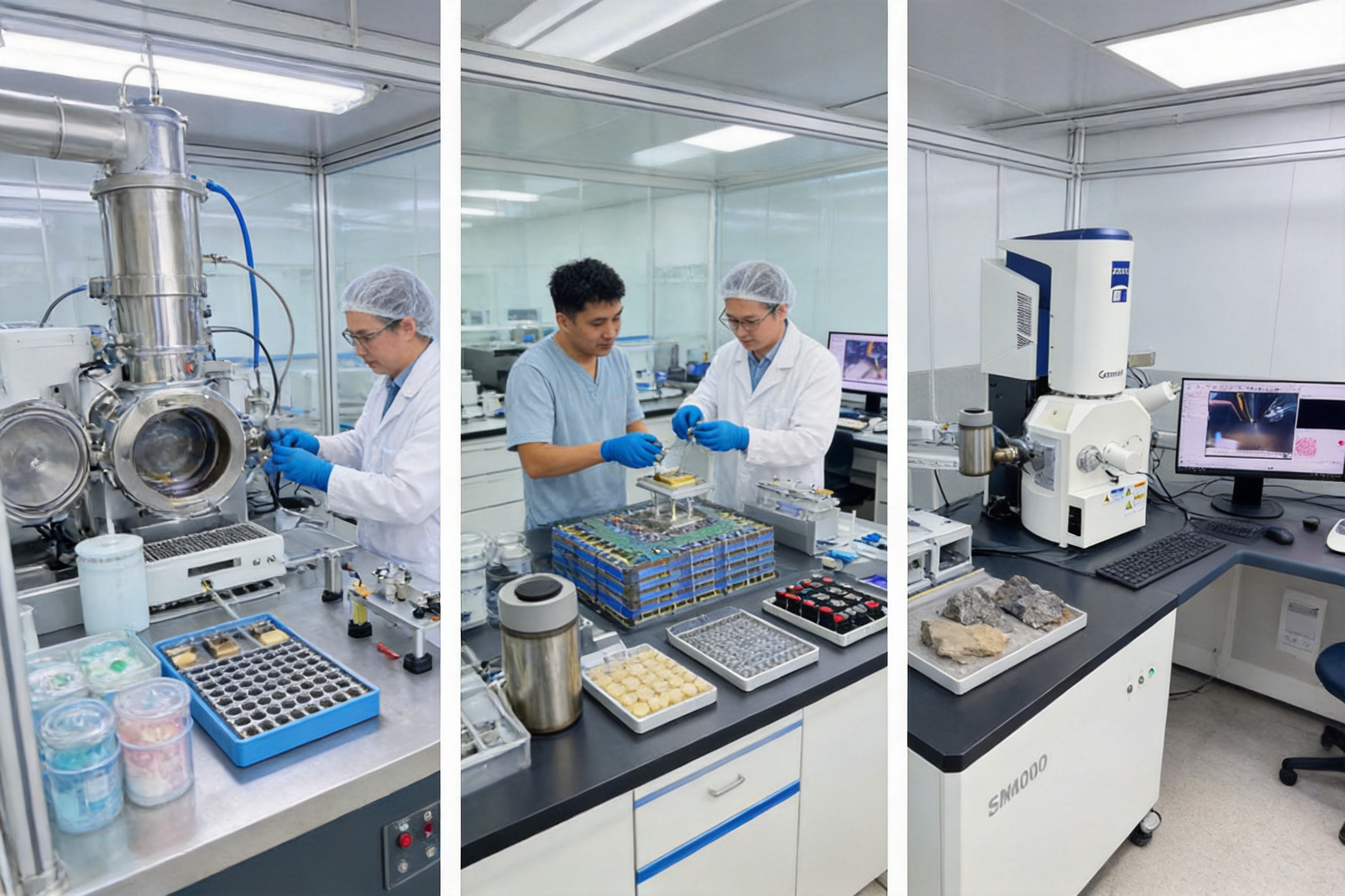
The performance of a sample preparation platform depends on the quality of its engineering and manufacturing process. Precision laboratory equipment must combine mechanical stability, vacuum compatibility, temperature control, electrical safety, process repeatability, and ease of operation. These requirements cannot be met through appearance or assembly alone.
The manufacturer behind this platform is a technology-driven enterprise with a dedicated research and development team focused on laboratory equipment, electronic testing, and safety compliance testing. Its development history began with an engineering studio specializing in electronic testing and later expanded into independently developed production lines and high-end laboratory equipment.
Research-Led Product Development
A research-led development model allows the product to be designed around actual laboratory problems rather than simple catalog specifications. Cryogenic transfer, ion milling, coating, and sample preparation involve complex interactions between temperature, vacuum, beam conditions, material properties, and operator procedures. Product development therefore requires continuous technical evaluation and application feedback.
The company’s engineering background supports the development of customized equipment and application-specific configurations. This is important because a biological research center may require different fixtures, transfer conditions, and controls from a semiconductor failure-analysis laboratory.
Precision Mechanical Design
Sample preparation equipment requires stable positioning and controlled movement. Mechanical components must maintain alignment during milling, transfer, coating, and docking. Unwanted vibration or movement can reduce the quality of a prepared surface or make it difficult to reproduce a process.
Precision design also affects the repeatability of sample positioning. A reliable holder and transfer interface help operators maintain orientation between preparation and observation. This is particularly important when a specific cross-section, interface, or region of interest must be located again inside the electron microscope.
Vacuum and Environmental Control
Vacuum systems must be designed to limit contamination, manage outgassing, and maintain stable operating conditions. Materials, seals, fittings, chambers, and internal surfaces all contribute to vacuum performance. For cryogenic equipment, the design must also consider condensation, thermal gradients, and the safe handling of cold components.
Environmental control is not limited to pressure. Temperature stability, cleanliness, coating conditions, and sample exposure time all influence the final result. A well-engineered system provides operators with a controlled environment in which process variables can be adjusted and documented.
Electrical and Safety Engineering
Ion beam systems and electronic laboratory instruments require careful attention to high-voltage operation, interlocks, grounding, cooling, and fault protection. Safety functions must be incorporated into the equipment architecture and supported by clear operating procedures.
The company’s experience in electronic testing and safety compliance supports a systematic approach to these requirements. Quality management and technical certification activities further contribute to product reliability and user confidence.
Customization and International Supply Capability
Many advanced laboratories require more than a standard configuration. They may need compatibility with existing microscopes, customized sample holders, special transfer interfaces, application-specific coatings, or integration with a larger laboratory workflow.
A combination of in-house engineering and international trade experience allows the manufacturer to support customized solutions for overseas partners. This can include technical communication, configuration review, documentation, shipment coordination, installation support, and application training.
Customization is especially valuable for institutions that are building shared electron microscopy centers. Instead of purchasing isolated equipment with incompatible interfaces, the laboratory can develop a coordinated preparation line matched to its microscopes, sample types, and operating procedures.
Application in Life Sciences and Cryo-Electron Microscopy
Life science research is one of the most demanding application areas for cryogenic sample preparation. Cells, tissues, viruses, liposomes, protein complexes, and other biological materials may lose their original organization if dehydrated, chemically altered, or exposed to excessive heat.
The platform supports workflows involving rapid cryo-fixation, cryogenic transfer, cryo-fracture, cryogenic polishing, and conductive coating. These steps can help researchers observe structures closer to their prepared hydrated state and reduce the artifacts associated with conventional preparation.
In cryo-electron tomography, the sample is examined from multiple angles and reconstructed into a three-dimensional volume. The quality of this reconstruction depends on the preservation of internal structures and the absence of excessive ice or contamination. A stable transfer pathway is therefore a fundamental part of the complete workflow.
Potential research areas include cellular ultrastructure, organelle organization, viral morphology, synaptic architecture, disease mechanisms, and protein assembly. Hospitals, biomedical research centers, national laboratories, and university microscopy facilities can use the platform to support both basic and translational research.
Application in Materials Science
Materials scientists often need to examine interfaces, defects, precipitates, pores, grain boundaries, and phase distributions. These features may be only a few nanometers or micrometers in size, and their interpretation depends on a clean and accurately prepared surface.
Ion milling and polishing can reveal the internal structure of metals, ceramics, composites, and coatings. For battery materials, the platform can support cross-section analysis of electrode particles, current collectors, separators, solid-state electrolytes, and solid-electrolyte interphase layers.
Battery materials may be reactive, porous, soft, or sensitive to air and moisture. Cryogenic preparation can help preserve unstable phases and reduce changes caused by warming. A prepared cross-section may then be transferred for electron imaging and elemental analysis with less risk of exposing the specimen to damaging conditions.
Polymer composites and soft materials also benefit from controlled preparation. Their interfaces may smear under mechanical polishing or deform under vacuum. Low-temperature processing can improve dimensional stability and preserve phase boundaries for microscopy.
Application in Semiconductors and Microelectronics
Semiconductor structures are increasingly complex, with multilayer architectures, nanoscale gate features, advanced packaging, and densely integrated interconnects. Failure analysis often depends on exposing a precise region without damaging adjacent structures.
Ion milling and etching can support cross-sectional preparation for FinFET structures, advanced logic devices, memory components, interconnects, dielectric layers, and packaging interfaces. The resulting surface can be examined for voids, delamination, cracks, contamination, electromigration, and process defects.
For industrial users, the main advantage is the combination of precision and repeatability. A controlled preparation process can help distinguish manufacturing defects from artifacts created during sample preparation. It also supports communication between process engineers, failure-analysis teams, and external testing laboratories.
The platform is suitable for semiconductor manufacturers, integrated circuit design companies, packaging specialists, electronics producers, and third-party analytical service providers. Its modular configuration allows users to scale the workflow according to production, research, or failure-analysis needs.
Application in Geology, Pharmaceuticals, and Energy Research
Geological samples often contain complex mineral associations, pores, inclusions, and fracture networks. Ion beam preparation can expose internal features while reducing mechanical damage. This is useful for shale pore structure analysis, mineral inclusion studies, and investigation of mineral interfaces.
In pharmaceutical and nanomedicine research, liposomes, nano-drug carriers, emulsions, and soft formulations can be highly sensitive to drying and beam exposure. Cryogenic preparation and transfer can help preserve morphology and distribution. Researchers can then examine particle size, aggregation, encapsulation-related structures, and interactions with surrounding materials.
Fuel cell catalysts, solid-state electrolytes, electrode interfaces, and other energy materials also require high-quality cross-sections. The platform supports the investigation of catalyst distribution, degradation, pore networks, interfacial reactions, and structural changes after cycling or environmental exposure.
Recommended Workflow for High-Fidelity Sample Preparation
A successful workflow begins with identifying the sample’s physical and chemical sensitivities. Operators should determine whether the specimen is hydrated, volatile, beam-sensitive, electrically insulating, mechanically fragile, reactive, or composed of multiple phases.
The next step is selecting the preparation temperature and environment. Stable metals and ceramics may be prepared under ambient conditions, while biological samples, polymers, electrolytes, and volatile materials may require cryogenic protection.
Bulk material can be reduced using an appropriate preliminary method, followed by ion milling or polishing to refine the analytical surface. The operator should select beam conditions that balance material removal rate, surface quality, heat generation, and possible ion-induced damage.
If the sample is nonconductive, coating may be applied to reduce charging. The coating thickness and material should be selected according to the imaging mode and analytical objective. For cryogenic samples, the coating step must be compatible with the required low-temperature condition.
Finally, the prepared specimen should be transferred using the appropriate LSCT configuration or compatible controlled pathway. The objective is to maintain orientation, temperature, cleanliness, and structural integrity until the sample reaches the electron microscope.
| Workflow stage | Primary consideration | Platform contribution |
| Sample assessment | Determine sensitivity to heat, vacuum, moisture, charging, and mechanical stress | Supports selection of ambient or cryogenic processing |
| Initial preparation | Reduce bulk material without compromising the region of interest | Provides a suitable starting geometry for ion processing |
| Precision milling or polishing | Remove damage and expose a clean surface or cross-section | IMP120 and HSC systems support controlled ion processing |
| Coating | Improve conductivity and imaging stability where required | HSC coating functions support application-specific surface preparation |
| Transfer | Prevent warming, contamination, and orientation loss | LSCT systems support cryogenic and vacuum-compatible transfer |
| Observation and analysis | Obtain representative images and analytical signals | Prepared samples are suitable for SEM, TEM, FIB, and cryo-EM workflows |
Operational and Commercial Benefits
In addition to technical performance, the platform offers practical benefits for organizations managing complex laboratory operations. Combining complementary functions can simplify equipment planning and reduce the need to coordinate unrelated vendors for every preparation stage.
A unified product family can also make operator training more efficient. Staff members can develop consistent procedures for sample handling, temperature control, ion processing, coating, and transfer. Shared documentation and common technical support can further improve facility management.
For service laboratories, a flexible platform expands the range of samples that can be accepted. The same facility may support biological research, materials characterization, semiconductor analysis, and energy-storage development. This diversification can improve instrument utilization and create additional value for customers.
For manufacturers, reliable sample preparation can shorten failure-analysis cycles and accelerate research and development. The ability to prepare a cross-section quickly and repeatably may help engineers identify process problems earlier, compare material formulations, and validate design changes.
Quality, Reliability, and Long-Term Development
High-end laboratory equipment must be reliable over many years because users often depend on it for irreplaceable research samples and time-sensitive industrial investigations. Reliability requires appropriate component selection, process inspection, assembly control, testing, and after-sales technical support.
The manufacturer has continued to improve its quality management system and technical specifications while expanding its product range. Its development path reflects a long-term emphasis on combining engineering capability with practical market and application requirements.
The company’s core philosophy, centered on precision craftsmanship and long-term innovation, is reflected in the platform’s intended use. The goal is not simply to supply an instrument, but to provide a dependable technical solution that can evolve with changing microscopy methods and sample types.
Future development opportunities include greater automation, digital process records, remote diagnostics, improved environmental sensing, recipe management, and closer integration with laboratory information systems. These improvements can support intelligent and data-driven sample preparation while preserving the operator’s ability to optimize parameters for unusual specimens.
Choosing the Right Configuration
When selecting a configuration, users should begin by defining the most important application requirements. A cryo-electron microscopy center may prioritize transfer temperature, vacuum compatibility, holder interfaces, and contamination control. A materials laboratory may focus on milling accuracy, surface finish, processing speed, and coating flexibility.
The sample volume and frequency of operation should also be considered. A research group processing a few specialized samples each week may require a flexible manual configuration. A high-throughput analytical center may benefit from standardized fixtures, repeatable recipes, and workflow optimization.
Compatibility with existing instruments is another key factor. The transfer system, sample holders, preparation chambers, and microscope interfaces should be reviewed before purchase. Technical consultation during the planning stage can help avoid compatibility issues and support future expansion.
Finally, users should evaluate technical support, training, documentation, spare parts, and customization capability. These factors have a direct effect on the total cost of ownership and the time required to achieve stable operation.
Why This Platform Is a Strong Competitive Choice
The competitive strength of the Electron Microscopy Sample Preparation Platform comes from the combination of complementary functions rather than from a single isolated specification. Cryogenic transfer protects sample state. Ion milling improves surface and cross-sectional quality. Coating and etching address conductivity, morphology, and localized preparation needs. Together, these capabilities cover a broader portion of the electron microscopy preparation workflow.
Compared with conventional mechanical preparation alone, the platform provides greater control over fragile interfaces and sensitive surfaces. Compared with disconnected equipment, it reduces unnecessary transfers and supports better process continuity. Compared with systems limited to either ambient or cryogenic operation, it offers greater application flexibility.
Its manufacturer also contributes important advantages through research-led engineering, customized design, electronic testing experience, quality-system development, and international supply capability. These strengths are valuable for customers who need more than a standard machine and require a complete technical solution suited to their laboratory environment.
For organizations working at the intersection of biology, materials science, semiconductors, energy, pharmaceuticals, and nanotechnology, the platform provides a practical foundation for high-fidelity electron microscopy. It helps laboratories protect the sample, refine the surface, control the transfer path, and obtain data that more accurately reflects the structure under investigation.
Questions and Answers
What is an electron microscopy sample preparation platform?
It is an integrated group of auxiliary instruments used to prepare, process, protect, transfer, and coat specimens before electron microscopy. The platform described here includes cryogenic transfer systems, multi-beam ion milling equipment, and ion beam etching, coating, and milling systems.
Which electron microscopes can use the platform?
The systems are intended to support workflows associated with scanning electron microscopes, transmission electron microscopes, focused ion beam instruments, cryo-electron microscopes, and related analytical platforms. Final compatibility should be confirmed according to the microscope model, sample holder, and transfer interface.
Why is cryogenic transfer important?
Cryogenic transfer helps protect frozen samples from warming, moisture, contamination, and ice crystallization. It is especially important for biological specimens, hydrated materials, volatile samples, and soft materials whose structure may change at room temperature.
Can hard materials be processed with the same platform?
Yes. Metals, ceramics, semiconductors, geological specimens, battery materials, and other hard materials can be processed through ambient ion milling, polishing, etching, and coating. The appropriate operating conditions depend on material composition, hardness, thickness, and analytical purpose.
What is the benefit of multi-beam ion milling?
Multi-beam capability provides greater flexibility in balancing removal rate, surface refinement, localized preparation, and temperature control. It allows the system to be adapted to different sample types and preparation objectives rather than relying on one fixed milling condition.
Can the platform process biological samples?
Yes. Biological samples such as cells, tissues, viruses, and protein-related structures can benefit from cryogenic fixation, cryogenic transfer, cryo-fracture, low-temperature milling, and controlled coating. These processes help preserve morphology and reduce artifacts caused by dehydration or warming.
Is coating always necessary?
No. Coating is application-dependent. It is often useful for nonconductive samples that experience charging during scanning electron microscopy, but coating thickness and material must be selected carefully when high-resolution imaging or chemical analysis is required.
How does the platform improve reproducibility?
It provides a more controlled sequence for preparation, processing, transfer, and observation. Operators can define procedures, manage environmental conditions, preserve sample orientation, and document process parameters. This can reduce variation between samples and users.
Can the equipment be customized?
Customized configurations may be developed according to sample type, microscope interface, transfer requirements, coating needs, laboratory layout, and application workflow. Technical evaluation is recommended before ordering to ensure that the proposed configuration matches the user’s existing equipment.
Who are the primary users?
Typical users include universities, national laboratories, electron microscopy centers, hospitals, biomedical research institutes, semiconductor companies, battery and energy-material manufacturers, pharmaceutical organizations, advanced-materials companies, and third-party testing providers.
What should customers consider before purchasing?
Customers should evaluate the sample types, required temperature range, preparation goals, microscope compatibility, throughput, vacuum requirements, coating options, operator skill level, maintenance needs, and technical support. A complete workflow assessment is more useful than selecting equipment based on one specification alone.
Conclusion
The quality of electron microscopy data begins with the quality of the sample. The Electron Microscopy Sample Preparation Platform addresses this fundamental requirement by combining cryogenic and vacuum transfer with precision ion beam processing, polishing, etching, milling, and coating.
LSCT-series systems help preserve frozen and temperature-sensitive specimens. The IMP120 supports accurate surface refinement and cross-section preparation under ambient or cryogenic conditions. HSC-series systems add flexible ion beam etching, coating, and milling functions. Used together, these technologies create a coordinated preparation ecosystem for biological research, materials science, semiconductor analysis, energy development, pharmaceuticals, geology, and nanotechnology.
The platform’s advantages over conventional or disconnected workflows include improved sample-state preservation, reduced mechanical damage, better contamination control, broader temperature flexibility, enhanced surface quality, and stronger process reproducibility. Supported by research-led product development, precision engineering, electronic testing expertise, quality management, and customization capability, it is positioned as a practical solution for laboratories that require reliable and high-fidelity electron microscopy preparation.
As electron microscopy continues to move toward higher resolution, three-dimensional reconstruction, cryogenic analysis, correlative methods, and automated interpretation, sample preparation will become even more important. A controlled, integrated, and adaptable platform gives researchers and industrial users a stronger foundation for obtaining accurate information from increasingly complex specimens.
References
1. Egerton, R. F. Physical Principles of Electron Microscopy: An Introduction to TEM, SEM, and AEM. Springer.
2. Williams, D. B., and Carter, C. B. Transmission Electron Microscopy: A Textbook for Materials Science. Springer.
3. Reimer, L., and Kohl, H. Transmission Electron Microscopy: Physics of Image Formation. Springer.
4. Frank, J. Three-Dimensional Electron Microscopy of Macromolecular Assemblies. Oxford University Press.
5. de Jonge, N., and Ross, F. M. Electron Microscopy of Specimens in Liquid. Nature Nanotechnology.
6. Echlin, P., Straw, M., Randolph, S., Filevich, J., and Kline, R. Ion Beam Preparation of Materials for Electron Microscopy. Materials characterization literature.
7. Goldstein, J., Newbury, D., Joy, D., Lyman, C., Echlin, P., Lifshin, E., Sawyer, L., and Michael, J. Scanning Electron Microscopy and X-Ray Microanalysis. Springer.
8. Committee on Advanced Electron Microscopy Practices. Guidelines for Specimen Preparation, Contamination Control, and Analytical Reproducibility. Technical reference literature.
Product: Electron Microscopy Sample Preparation Platform
PREV:Advanced Ion Beam Etching and Thin Film Deposition for Precision Materials Processing
NEXT:No next article
NEXT:No next article
Interested in cooperation or have questions?
Featured Products
Related products
Quick Link
Contact Information
- Tel: +86-13861043568
- E-mail: 1840784112@qq.com
- Add: Tianting District, Changzhou City, Jiangsu Province, China, No. 6 Hecircuit
Scan Code
Copyright © JIANGSU BAISHENG INDUSTRIAL CO., LTD.
All Rights Reserved. Privacy Custom Precision Laboratory Testing Equipment Manufacturers
